Research
Research Overview
Our research involves developing a fundamental understanding of the chemical, photophysical, and optical properties of nanometer scale materials. Unlike macroscopic materials, these objects have physical characteristics that are strong functions of their size and shape. Thus, nanometer scale materials have properties that can be easily controlled and manipulated. Currently, our investigations are focused on the synthesis and spectroscopic characterization of the chemical and physical properties of carbon nanotubes and semiconductor nanocrystals (NCs), and the application of these materials to solving important challenges in energy, photo-catalysis, quantum optics, and biomedicine. These studies are highly interdisciplinary, and lie at the interface between chemistry, physics, applied physics, and materials science.
Topics
Materials | Experimental Techniques | Current ResearchMaterials
A. Quantum Dots
Colloidal semiconductor NCs are an important class of material due to their rich diversity in structure and optical properties. Precise control over the morphology, and subsequently optical properties, of these materials have afforded solution-processed nanomaterials a breadth of applications in lasing, bioimaging, solar light concentration, and more.
Our laboratory is currently involved in many projects utilizing colloidal quantum dots (QDs). We are part of a collaborative effort focused on using QDs as photo-redox catalysts in organic reactions. By probing mechanistic details and investigating the potential reactions that QDs can favorably catalyze, this work showcases how the unique properties of QDs can be used to redefine chemical reactivity. We also study the use of QD-sensitized photocathodes for hydrogen production. Fabrication of QD photocathodes via deliberate synthetic and engineering techniques will enable the efficient shuttling of electrons and holes from the electrode to the solution.

(Left) General scheme detailing the use of colloidal QDs as photo-redox catalysts in organic reactions. (Right) A rainbow photocathode (PNAS, 2017, 114, 43, 11297-11302) as well as the corresponding energy levels of different-sized CdSe QDs and the possible electron transfer process between them.
B. Nanoplatelets
Recent advancements in the synthesis of colloidal semiconductor NCs have produced atomically flat NCs known as nanoplatelets (NPLs). NPLs exhibit quantum confinement in only one crystal direction which leads to strong dependence of the emission and absorption features on the thickness of the NPL. This NC dimension can be synthetically controlled with monolayer precision producing homogeneous samples and narrow ensemble linewidths which are essential for applications in lasing and LEDs.

(Left) TEM micrograph of 4 monolayer CdSe NPLs. The scale bar defines a 50 nm length. (Right) Optical properties of 3, 4 and 5 monolayer NPLs. Photoluminescence emission shifts to longer wavelengths as the number of monolayers increases.
Our research surrounding NPLs focuses primarily on the cadmium chalcogenides which are currently of great interest in the NC community. Our group is particularly interested in the optical and electronic properties of NPLs with various thicknesses, lateral areas, and chemical compositions. Specifically, we are interested in examining the binding nature and electronic effects of heterovalent dopant ions that are incorporated into the NPLs. These dopant ions can localize excited charge carriers, which results in unique optical and chemical properties.In addition, our group is working to combine concepts from natural photosynthesis with the attractive photosensitizing properties of NPLs for renewable energy applications, such as hydrogen production via water splitting.

(Left) Ensemble UV/Vis absorbance and PL spectra of undoped (green) 4.5 ML CdSe NPLs & Ag-doped (orange) NPLs leading to red-shifted, broad dopant PL emission arising from hole-trapping Ag+ sites within the lattice (inset). (Right) Single particle characterization of Ag:CdSe NPLs reveal dual emission from both band-edge and dopant recombination with spectral diffusion shown (inset) for a single NPL.
C. Single-walled Carbon Nanotubes
Single-walled carbon nanotubes (SWCNTs) have remarkable properties and many potential applications in different fields of both science and technology. A SWCNT consists of a hexagonal network of carbon atoms (a graphene sheet) rolled up into a cylinder with a diameter typically less than 1 nm and a length on the order of a few to hundreds of microns. Unique properties of the carbon-carbon bond make SWCNTs both extremely strong and flexible. Interestingly, SWCNTs can have either metallic or semiconducting properties depending on their diameter and helicity. While metallic nanotubes are very good electron field emitters, semiconducting SWNTs offer stable and tunable fluorescence in the near-infrared region (NIR). SWCNTs are potentially useful for applications in diverse areas such as molecular electronics, biotechnology and bioimaging, energy storage, and quantum information sciences. We have several projects centered around studies of SWCNT photophysical and charge properties using diverse spectrometric and microscopic methods.

(Left) Schematic depicting the formation of a SWCNT from a rolled up sheet of graphene (Acc. Chem. Res. 2008, 41, 2, 235–243). (Right) . Depiction of a SWCNT. Charge carriers are able to freely propagate down the length of the SWCNT (blue arrows), but are circumferentially confined due to the nanometer size scale.
Experimental Techniques
A. Time-Resolved Setup: Single Photon Measurements
Single photon sources that will be key building blocks towards optical based quantum information science applications will need to demonstrate both high photon purity and indistinguishability. Using a confocal microscope, we measure these single emitter (i.e., QDs, SWCNTs w/ defects, etc.) properties by characterizing its (1) fluorescence lifetime, (2) quantum coherence (Hong-Ou-Mandel (HOM) two photon interference), and (3) photon antibunching. A Hanbury Brown & Twiss interferometer with single-mode polarization maintaining fiber delay lines is used to measure the second order photon correlation function, g(2)(t), and extract the two photon interference visibility along with photon decoherence timescales. Measurements taken as a function of temperature will also determine the extent of any exciton-phonon coupling on dephasing mechanisms in various NC systems.

(Left) PL spectra and blinking time trace of individual gradient core-shell CdSe/CdS QDs. (Right) Single photon emission from QDs via pulsed-laser excitation leading to photon antibunching under ambient conditions.
B. Ultrafast Setup: Charge Transfer Measurements
Ultrafast spectroscopy techniques (pump-probe spectroscopy) use sequences of ultrashort light pulses (with typically femtosecond durations) to study photoinduced dynamical processes in atoms, molecules, nanostructures, and solids. Our group is interested in studying the charge transfer dynamics in semiconductor NCs in systems relevant for hydrogen production. Our current setup allows us to perform single-color pump-probe experiments whilst YAG-generated white light allows us to probe dynamics up to 1000 nm. Ongoing projects are focused on studying the hole transfer/extracellular electron transfer processes in systems involving CdSe QDs and polyoxovanadate clusters/Shewanella oneidensis respectively. Recently, we are developing our setup to enable the study of ultrafast population dynamics in cavity polariton systems.

C. Scanning Probe Microscopy
Using electrostatic force microscopy (EFM), a variation of atomic force microscopy, we quantitatively determine localized charges on nanomaterials at the nanometer scale. In EFM, a conductive AFM tip is lifted off the substrate and a voltage is applied between the oscillating tip and a grounded sample. The cantilever response due to long-range electrostatic forces is then measured as it is raster scanned across the surface.

Simplified Schematic of EFM experimental setup.
The impact of the local environment on individual nanomaterials can be further investigated by correlating single molecule spectroscopy (SMS) with atomic force microscopy (AFM), which allows our laboratory to study the optical response of nanomaterials to physical perturbations.

Simplified Schematic of Correlated AFM-SMS setup.
Current Research
A. Photocatalysis and Hydrogen Generation
Global energy demands and climate crises have made carbon-free energy a growing area of research interest. The use of hydrogen molecules as such a fuel is a promising due to its high energy density and clean usage, however, the challenges lie in production of hydrogen from sustainable sources. Colloidal semiconductor NCs have shown to be useful tools in the production of hydrogen through photocatalysis.
Our laboratory is currently involved in two collaborative projects focusing on hydrogen generation via photocatalysis. These projects involve using QDs and NPLs as photosensitizers to harvest photons and subsequently supply electrons for proton reduction. These two projects utilize different systems:
1. Nanoscale and Biomimetic Assembles
Our nanoscale photocatalysis systems take inspiration from natural photosynthesis. Our QDs and NPLs harvest light and accumulate charge, then transfer the charge to reduce protons, yielding gaseous hydrogen. In this system, a co-catalyst and sacrificial electron donor help facilitate hydrogen generation. Our lab is currently trying to understand fundamental questions within these photocatalytic systems, studying the effect different crystal structures and capping ligands have on the catalytic activity of QDs as well as where catalysis takes place on a NPL by implementing different heterostructures (core-crown and core-shell NPLs).

(Left) General scheme demonstrating hydrogen generation using QDs, sacrificial electron donor, and co-catalyst (J. Chem. Phys.. 2021;154(3). doi:10.1063/5.0032172). (Right) General scheme demonstrating hydrogen generation using NPLs, sacrificial electron donor, and co-catalyst.
2. Living Bio-Nano Systems
Our study of hydrogen production in living bio-nano systems uses Shewanella oneidensis MR-1, naturally occurring bacteria in the lakes near Rochester, as sustainable electron donors to facilitate the hole-transfer process in the photoexcited QDs. The photoexcited electron from the conduction band then reduces the protons in the medium leading to sustainable hydrogen production via hydrogen evolution reaction (HER). This bio-nano assembly overcomes the limitations of molecular electron donors and oxygen evolution reaction (OER) by sustainable electron generation through lactate consumption by bacteria.
Our lab is interested in studying the effects of different capping ligands, compositions, morphologies, and sizes of nanocrystals in this assembly. The bio-nano interface through which the extracellular electron transfer (EET) from bacteria to photocatalysts occurs is probed by correlated atomic force microscopy (AFM), NMR, FTIR, fluorescence, transient absorption spectroscopy, gas chromatography, and transmission electron microscope imaging.

General scheme detailing the photocatalytic system using QDs and bacteria (PNAS, 2023, 120, 17, e2206975120, doi.org/10.1073/pnas.2206975120).
B. Optimization of Synthetic Parameters Using Artifical Intelligence
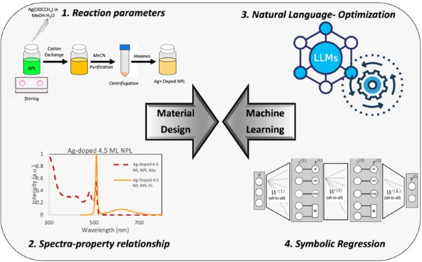
Artificial intelligence (AI) enables the merger of computational tools and traditional material synthesis. Especially, the use of large natural language models (LLMs) and symbolic regressions (SR) to mimic human cognitive analysis and to build formulaic relationships is advancing the field of material design and analysis. Our current work, in collaboration with the White lab, focuses on the AI-enabled precise post-synthetic Ag+ doping of CdSe NPLs and their spectroscopic analysis.

C. Investigating Colloidal Semiconductor Nanoplatelet Charge Characteristics
Due to ligand passivation, the as-synthesized NPLs are nominally charge neutral, however, the charge characteristics are greatly altered if dopant atoms are introduced into the lattice. Comparing the charge before and after doping provides insights into the way the dopant enters the lattice. For 4.5 monolayer CdSe NPLs doped with silver, we observe a significant decrease in charge suggesting that the silver is substitutionally doped in the lattice.

D. Investigating Single-Walled Carbon Nanotube Photophysics and Charge Characteristics
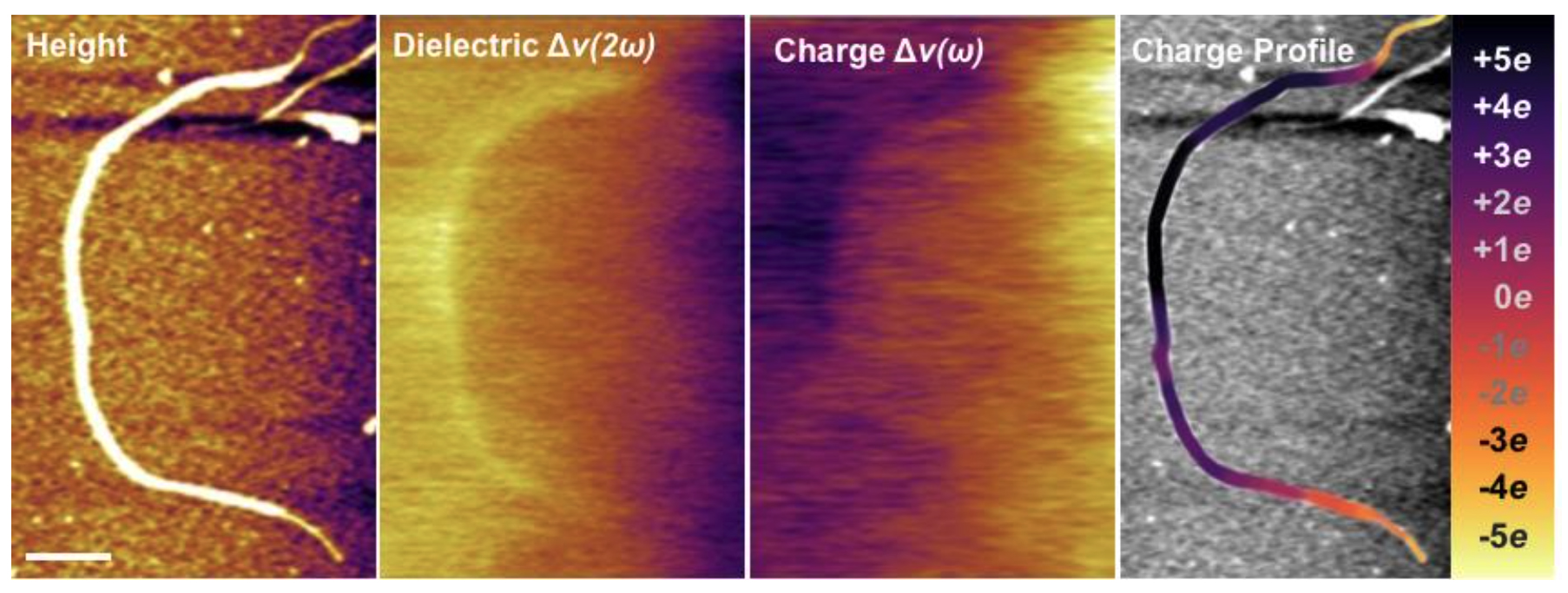
As-synthesized, SWCNTs are nominally charge-neutral, however, ionic surfactants that are commonly used to disperse SWCNTs in solution can lead to heterogeneous surface charge buildup along the tube. For dozens of long SWCNTs dispersed with ionic surfactant, we found surprisingly large charge density variations all along the tube. Acquiring EFM data from before and after photoexcitation suggests that the ionic surfactant creates local electrostatic perturbations that act as barriers for exciton transport, which leads to distinct localized regions of charge.

EFM images of (from left to right) topography, dielectric constant, charge and charge profile for an individual SWCNT covered in surfactant. The inset is the charge magnitude scale ranging from -5eto +5e. Dark (bright) contrast in the charge image is positive (negative) charge. Scale bar is 200 nm.
We discovered that regions of individual SWCNTs covered in surfactant aggregates display localized and redshifted PL character. From our EFM measurements, we know that these same regions routinely display greater static charge accumulation. Together, the correlated SMS-AFM measurements and EFM data suggests a strong connection between electrostatic and optical characteristics of SWCNTs.

(Left) SMS images overlaying AFM images. (Right) Emission energy profile for the individual SWCNT in the left panel. Two regions of intense surfactant aggregation demonstrate localized and redshifted PL emission.
E. Biological Imaging
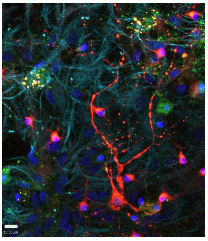
Compared to traditional fluorophores, colloidal semiconductor QDs exhibit enhanced photophysical properties that are of interest to biological applications, not limited to biosensing and bioimaging. Our previous work specifically elucidated which of these photophysical properties are essential to enhancing the localization precision of single-molecule super-resolution microscopy methods. Yet, despite the potential for QDs to improve the current limits of biophysical measurements, their adoption in biomedical investigations is limited. A key limitation of the adoption of QDs arises from concerns regarding steric hinderances due to the large size of QDs compared to traditional fluorophores. This ultimately necessitates additional control experiments to assess that QD-conjugated targets function similarly to their endogenous and small fluorophore-labeled counterparts.

However, rather than see the larger size of QDs as a detriment, our work is interested in capitalizing on this feature to construct fluorescent biomimetic structures of biological macromolecules. Our initial work focused on constructing biomimetic QDs for oligomeric amyloid-beta 42 to interrogate the specific subcellular interactions in the central nervous system(CNS)relevant to Alzheimer’s Disease. More recently, we have pivoted this work to construct biomimetic structures for SARS-CoV-2 virus particles, termed COVID-QDs, to elucidate how neurological dysfunction manifests in COVID-19 pathogenesis. The COVID-QDs serve as a proxy to interrogate what interactions the native virus may have in the CNSthat we will be able to observe by taking advantage of the superior optical properties of the QDs.

F. Quantum Information Science and Quantum Optics
Strong coupling between electronic states of molecules and photons confined within a cavity generates exciton-polaritons. These exciton-polaritons offer a platform to study nanoscale light-matter interactions and have gained significant attention due to their immense research prospects. Coupling molecules to a quantized radiation field inside an optical cavity has the potential to control photochemical reactivities and open new chemical reaction pathways. Our research focuses on characterising the photophysical and photochemical properties of cavity polaritons and exploring their potential applications.
The large oscillator strength of NPLs with highly symmetric in-plane optical transition dipole and their exceptionally narrow fluorescence linewidth make these semiconducting NCs a promising candidate for achieving strong light−matter coupling. We have recently utilized these NPLs as an active material in metal-DBR cavities to generate room-temperature exciton-polaritons. Several key findings include the largely temperature and detuning independence of Rabi splitting, the effect of cavity quality (Q) factor on excited state dynamics leading to upconversion, and the observation of polariton lifetimes on the order of 100 picoseconds, indicating their potential to affect photochemical reaction rates.

Angle-resolved reflectance (a,c) and PL (b,d) spectra with similar ΔE and ħΩR at T= 300 K (a,b) and T= 5k (c,d).

(Left) Angle-resolved reflectance & (Middle) PL spectra (experimental left, simulated right) with Δ= -1 meV, ħΩR= 31 meV and Q= 300. Quantum dynamical simulations show excellent agreement with measured spectra with white arrows indicating UPB PL emission. (Right) Theoretical calculations of the Upper Polariton population buildup with increasing cavity Q-factor.
Additionally, we are interested in investigating the effect that strong-coupling has on chiral light-matter interactions – interactions arising from either an optical field (cavity photon), exciton, or both exhibiting symmetries such that mirror reflection produces a change in spatial conformation (circular polarized light and a carbon with 4 different ligands attached satisfy this condition). Preserving chiral information in an optical cavity is challenging, requiring innovation in cavity design. Current thoughts in achieving this cavity symmetry include utilizing CdSe NPLs with chiral ligands demonstrating circular dichroism as the active material coupled to a Fabry-Perot cavity constructed with metasurface mirrors or a 2-D chiral active/spacer layer.

(Left) Absorbance, photoluminescence, and circular dichroism spectra of CdSe NPL film with chiral ligands. (Right) Metasurface mirror and associated reflection spectra upon illumination with different light polarizations. Adapted from Light Sci Appl 9, 23 (2020). https://doi.org/10.1038/s41377-020-0256-5.
Successful chiral strong-coupling could lead to an energy difference between chemical enantiomers, paving the way towards selective chiral synthesis, detection, or an increase in the degrees of freedom of a quantum communications platform.